Medical Disclaimer
  • For Educational Purposes Only: This content is intended for educational reference and should not be used for clinical decision-making.
  • Not a Substitute for Professional Judgment: Always consult your local protocols, institutional guidelines, and supervising physicians.
  • Verify Before Acting: Users are responsible for verifying information through authoritative sources before any clinical application.
AI Assistance Notice
The clinical content and references are curated and reviewed by myself; however, AI was used to assist in organizing, paraphrasing, and formatting the information presented.
Bedside Snapshot
Purpose
Home-based RRT using peritoneal membrane for solute/fluid removal via osmotic and diffusive exchange
Types
  • CAPD: Manual exchanges (4-5/day)
  • APD: Automated overnight cycler
Advantages
  • Home-based, patient independence
  • Gentler fluid removal
  • Preserves residual kidney function
Key Complication
  • Peritonitis (cloudy effluent, abdominal pain)
  • Exit-site/tunnel infection
What is Peritoneal Dialysis?

Peritoneal dialysis (PD) is a form of renal replacement therapy that uses the patient's own peritoneal membrane as the semipermeable "dialyzer" to remove waste products, correct electrolytes and acid-base disturbances, and remove excess fluid.

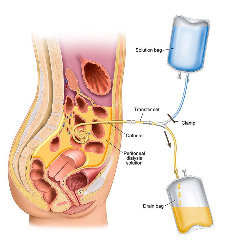
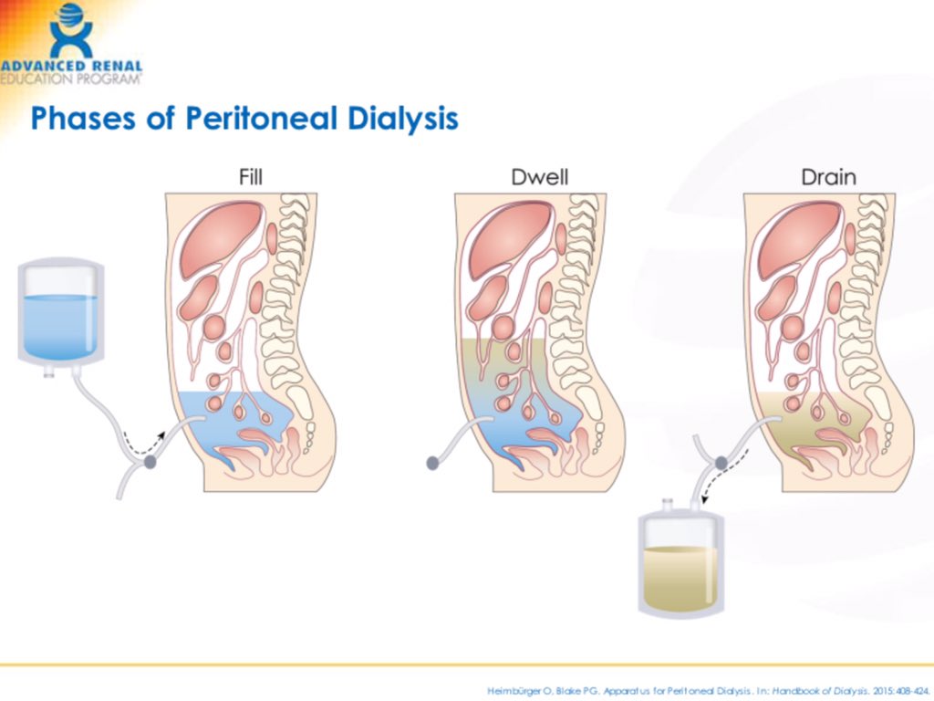
A soft catheter is placed into the peritoneal cavity. Sterile dialysis solution (dialysate) is infused into the abdomen, where it dwells in contact with the peritoneal capillary bed. Solutes and water move between blood and dialysate, and then the used fluid is drained out and replaced with fresh solution.

Basic peritoneal dialysis diagram showing the peritoneal cavity, PD catheter, dialysate in the abdomen, and blood vessels in the peritoneum
Figure 1: Peritoneal dialysis overview - catheter placement and dialysate in peritoneal cavity

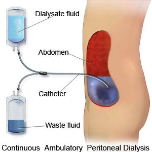
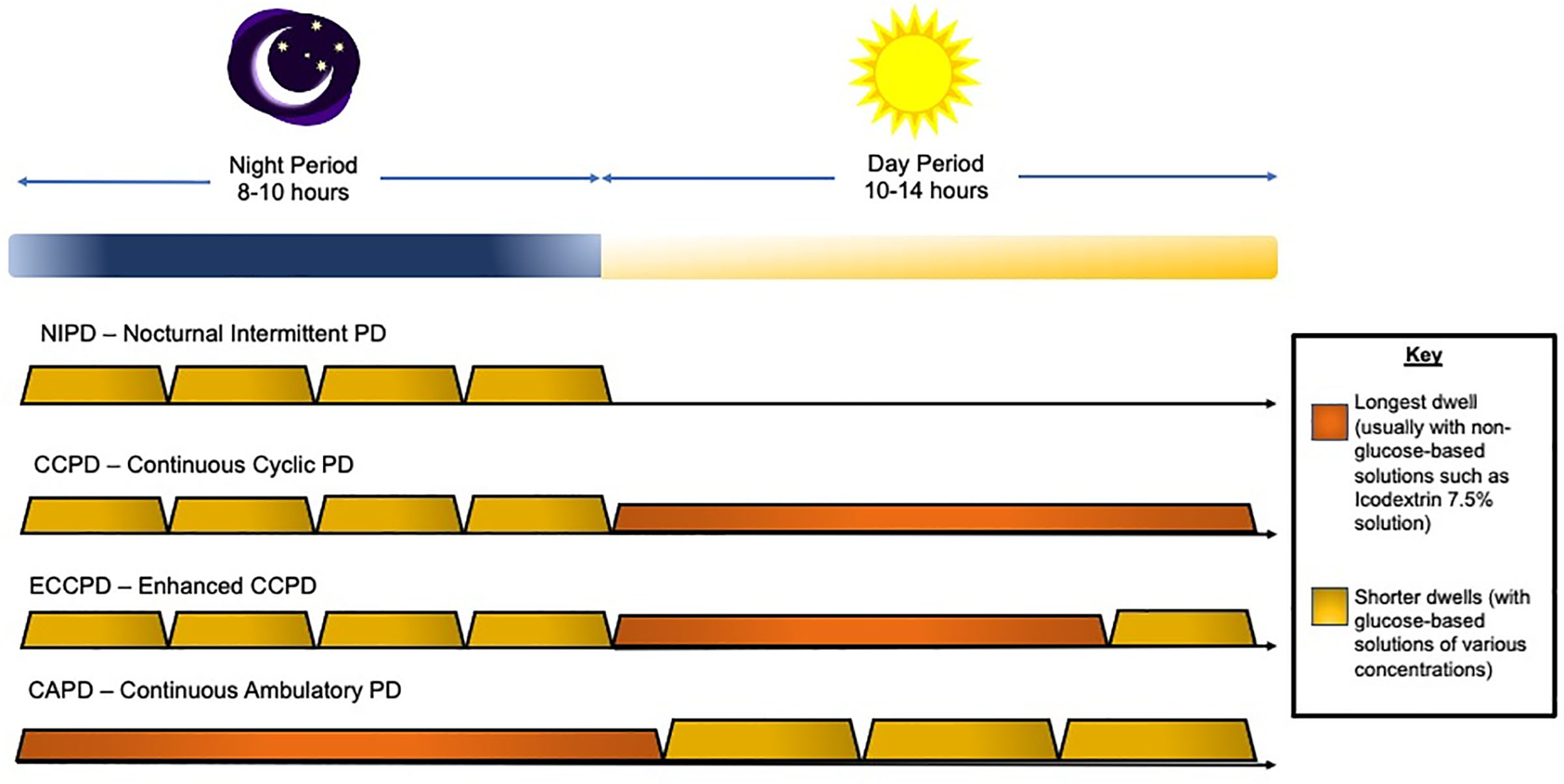
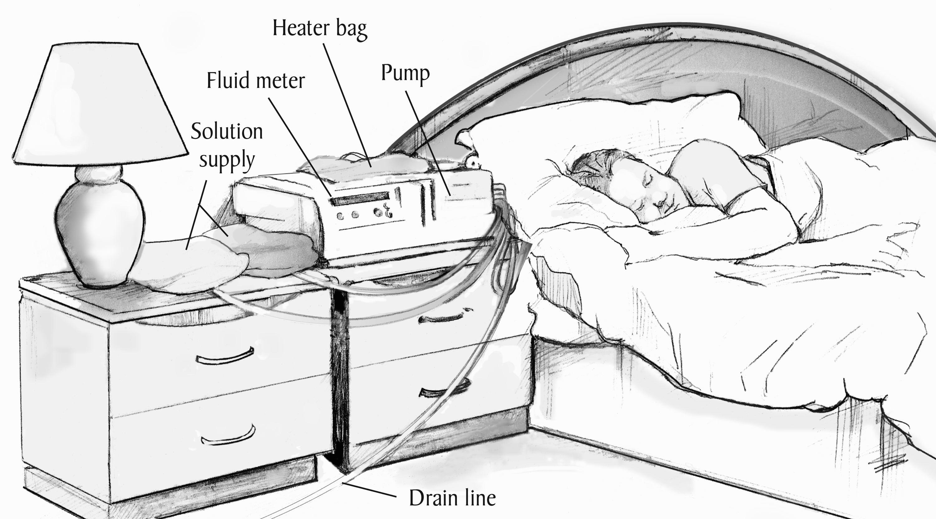
PD is most commonly a home-based therapy for patients with end-stage kidney disease (ESKD). It can be performed manually during the day (CAPD) or automatically at night using a cycler (APD). Nurses in acute and chronic settings support patient training, monitoring, troubleshooting, and infection prevention.

Types of Peritoneal Dialysis

There are two main types of peritoneal dialysis used in practice:

Continuous Ambulatory Peritoneal Dialysis (CAPD)

  • Manual exchanges performed by the patient or caregiver, typically 3–5 times per day
  • Each exchange involves draining the used dialysate, infusing fresh solution, and allowing it to dwell for several hours
  • No machine is required; treatment is continuous throughout the day

Automated Peritoneal Dialysis (APD) – also called CCPD

  • A cycler machine performs multiple exchanges overnight while the patient sleeps
  • The patient may wake up "full" (with dialysate dwelling) or "dry" depending on the prescription
  • APD can offer more flexibility for school, work, and daytime activities
Infographic comparing CAPD and APD including number of exchanges, timing, cycler requirements, and lifestyle considerations
Figure 2: CAPD vs APD comparison - timing, exchanges, and lifestyle fit
Automated peritoneal dialysis cycler with patient connected at bedside
Figure 3: APD cycler machine - automated exchanges performed overnight
Indications and Patient Selection

Peritoneal dialysis is one of several options for long-term dialysis in ESKD. It may be chosen for:

  • Patients who value home-based, flexible therapy and wish to avoid thrice-weekly in-center HD
  • Patients with limited vascular access options or severe vascular disease
  • Children and smaller adults where PD can be technically easier and preserve vascular access for the future
  • Patients who live far from a dialysis center or have transportation barriers

Selection Considerations

  • Ability to perform or supervise sterile exchanges (patient or caregiver), including adequate vision, dexterity, and cognition
  • Suitable home environment: clean area for exchanges, storage space for supplies, reliable electricity (especially for APD)
  • Medical factors: residual kidney function, abdominal anatomy, hernias, prior major abdominal surgeries, obesity, and comorbidities
Contraindications and Limitations

Absolute or strong contraindications are uncommon but include situations where PD cannot function or would be unsafe.

Examples (often relative and case-dependent)

  • Extensive intra-abdominal adhesions or scarring severely limiting dialysate distribution
  • Active abdominal wall or intra-abdominal infection (e.g., peritonitis, diverticulitis) until resolved
  • Large non-reducible abdominal hernias or severe diaphragmatic defects not corrected surgically
  • Severe protein loss or malnutrition that would be worsened by PD
  • Inability to safely perform sterile technique at home with no available support

Limitations Compared with Hemodialysis

  • Clearance may be less efficient in very large patients or those with high catabolic rates
  • Glucose-based solutions can contribute to weight gain, hyperglycemia, and dyslipidemia
  • Risk of peritonitis, exit-site, and tunnel infections
  • Long-term risk of peritoneal membrane changes (e.g., fibrosis, encapsulating peritoneal sclerosis) with many years of PD
Peritoneal Dialysis Catheter and Access

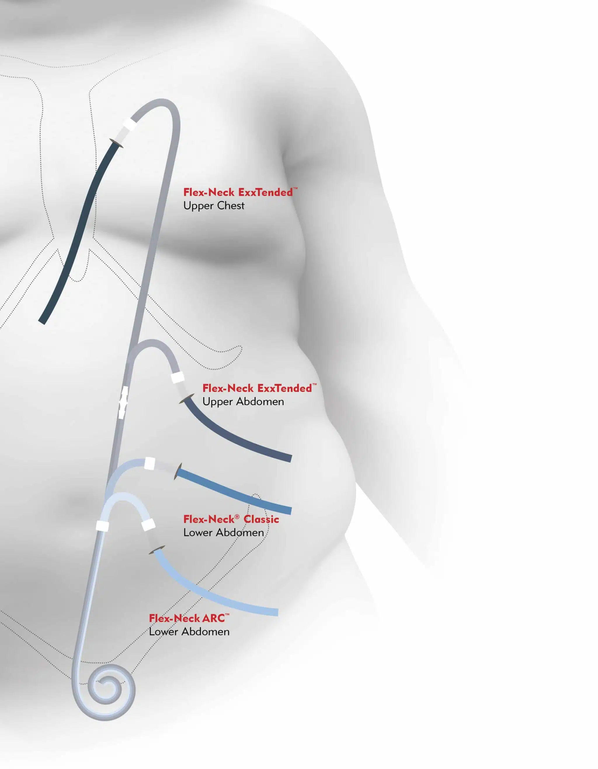
The PD catheter is a soft, flexible tube placed into the peritoneal cavity, usually via laparoscopic or open surgical technique.

Key Features

  • The intraperitoneal segment lies in the pelvis, often with curled or coiled tips to reduce migration
  • One or two Dacron cuffs sit within the subcutaneous tunnel to anchor the catheter and reduce infection risk
  • The exit site is brought out through the abdominal wall where daily cleaning and dressing changes occur

Common Exit Site Locations

  • Lower abdomen (below or lateral to the umbilicus), directed downward to reduce contamination and avoid beltlines
  • Extended or presternal catheters for patients with obesity, ostomies, or other abdominal issues
Illustration of PD catheter positions and exit-site options including standard lateral, downward exit, and presternal catheter
Figure 4: PD catheter positions and exit-site options
Patient education diagram showing abdominal PD catheter placement and exit site
Figure 5: Patient education - PD catheter placement and exit site care

Nursing Considerations

  • Inspect the exit site every shift (in hospital) and at routine clinic visits for redness, drainage, pain, and crusting
  • Teach patients the importance of daily exit-site care and keeping the catheter secure and dry
  • Reinforce that the catheter is for PD only—not for infusions, blood draws, or medications
Critical Rule: The PD catheter is for peritoneal dialysis ONLY. Never use for IV access, blood draws, or medication administration.
Principles of PD and Basic Prescription

PD works through diffusion and osmosis across the peritoneal membrane:

  • Diffusion: Small solutes (urea, creatinine, potassium) move down their concentration gradient from blood to dialysate
  • Osmosis/ultrafiltration: Glucose in the dialysate creates an osmotic gradient that pulls water out of the blood into the dialysate

Basic Elements of a PD Prescription

  • Type of PD: CAPD vs APD
  • Fill volume per exchange: 1.5–3.0 liters per dwell in adults, adjusted for body size and tolerance
  • Number of exchanges: Per day (CAPD) or per night (APD)
  • Dwell time: How long fluid remains in abdomen (commonly 4–6 hours for daytime CAPD; shorter cycles at night on APD)

Dialysate Strength and Composition

  • Glucose concentration (1.5%, 2.5%, 4.25%) to adjust ultrafiltration
  • Icodextrin solutions for long dwells to reduce glucose exposure and improve UF
  • Calcium and magnesium content tailored to labs and bone-mineral management
Three steps in peritoneal dialysis exchange: Drain, Fill, Dwell cycle diagram
Figure 6: PD Exchange Cycle - Drain → Fill → Dwell (Source: NIDDK)
Clearance Monitoring: Kt/V and clinical assessment (symptoms, volume status, labs) are used by nephrology to evaluate PD adequacy.
CAPD Exchange Workflow (Manual PD)

A typical CAPD exchange (manual PD) involves a structured, aseptic process. Details vary by system (Baxter, Fresenius, etc.), but the core steps are similar:

Video: CAPD Exchange Procedure Demonstration (~7 minutes)
  1. Prepare:
    • Perform hand hygiene, wear mask as per local protocol, and set up supplies in a clean, well-lit area
    • Check solution bags (clarity, expiration date, correct glucose concentration, and temperature if pre-warmed)
    • Inspect catheter and dressing before starting
  2. Connect:
    • Don mask and perform hand hygiene again
    • Connect the PD catheter to the transfer set/system tubing using aseptic technique
  3. Drain:
    • Open the appropriate clamp to allow used dialysate to drain from abdomen into drain bag
    • Observe effluent for clarity, color, fibrin strands, and volume
  4. Flush (if applicable):
    • Flush a small amount of fresh dialysate to clear the line and reduce contamination risk
  5. Fill:
    • Infuse the prescribed volume of fresh dialysate into the abdomen
    • Clamp lines and disconnect per system protocol
  6. Dwell:
    • Patient goes about normal activities while dialysate dwells for the prescribed time
  7. Disconnect and Secure:
    • Ensure catheter is clamped, capped, and secured
    • Perform final hand hygiene and dispose of used supplies appropriately
Routine Nursing Monitoring and Care

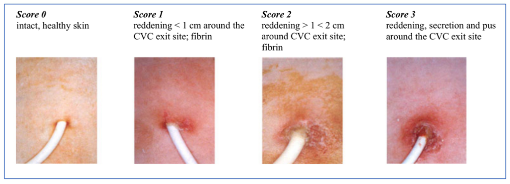
Key assessments for patients on PD (inpatient or outpatient visits):

Vital Signs and Weight

  • Vitals: Blood pressure, heart rate, respiratory rate, SpO₂, temperature
  • Weight: Track pre- and post-exchange or daily weight to monitor volume status and UF effectiveness

Intake/Output and Effluent

  • Net ultrafiltration (UF): Per exchange or per day; report unusually low or negative UF
  • PD effluent: Assess clarity, color, fibrin, and presence of blood or cloudiness

Physical Assessment

  • Abdomen: Evaluate for pain, distension, guarding, rebound, and hernias
  • Exit site and tunnel: Inspect for redness, drainage, swelling, tenderness, crusting, or overgranulation

Laboratory Monitoring

  • Electrolytes, BUN/creatinine, bicarbonate, calcium, phosphate, albumin, and blood counts (per schedule)
Documentation Checklist: Type and volume of dialysate, dwell times, UF volume, any complications during exchanges, and patient tolerance.
Common Complications of Peritoneal Dialysis

Peritonitis

  • The most serious PD-specific complication; usually presents with abdominal pain, cloudy effluent, and sometimes fever or GI symptoms
  • Diagnostic criteria: Cloudy effluent with elevated WBC count (e.g., >100 WBC/µL with ≥50% neutrophils) and/or positive culture, per ISPD guidelines
  • Management: Intraperitoneal antibiotics directed by local protocols and culture results; may require hospitalization in severe cases
Peritonitis Alert: Cloudy effluent + abdominal pain = suspect peritonitis until proven otherwise. Notify physician immediately and obtain effluent sample for cell count and culture.
Algorithm for diagnosis and management of PD-associated peritonitis based on ISPD guidelines
Figure 8: PD Peritonitis - Diagnosis and Management Algorithm (ISPD Guidelines)

Exit-Site and Tunnel Infections

  • Localized erythema, tenderness, or purulent drainage at catheter exit or along tunnel track
  • Increase risk of peritonitis; require prompt evaluation and antibiotic therapy per guideline

Mechanical Complications

  • Hernias: Umbilical, inguinal, or incisional due to increased intra-abdominal pressure
  • Leaks: Around the catheter or into abdominal wall/scrotum
  • Catheter malposition: Causing poor drainage or pain

Metabolic Complications

  • Hyperglycemia: Due to glucose absorption from dialysate
  • Weight gain and dyslipidemia: From chronic glucose load
  • Protein loss: In dialysate contributing to hypoalbuminemia and malnutrition

Other Complications

  • Back pain or discomfort from dwell volumes
  • Encapsulating peritoneal sclerosis (EPS): Rare, severe long-term complication after many years of PD
Infection Prevention and Exit-Site Care

Nursing and patient practices that reduce infection risk:

Aseptic Technique

  • Strict hand hygiene and mask use (per local protocol) during connections and disconnections
  • Using aseptic technique and closed systems during exchanges

Daily Exit-Site Care

  • Cleanse with recommended solution (e.g., antibacterial soap or chlorhexidine) as per program protocol
  • Apply topical antibiotic ointment if ordered (e.g., mupirocin or gentamicin) to reduce exit-site infection and peritonitis risk
  • Keep the site dry; avoid soaking baths or pools unless specifically cleared
Exit site care procedure and assessment guide
Figure 9: Exit Site Assessment and Care Guidelines

Additional Prevention Measures

  • Securing the catheter to prevent traction or trauma to the exit site
  • Education on promptly reporting any signs of infection: redness, increased pain, drainage, fever, or cloudy effluent
Prevention Pearl: Exit-site infections can lead to tunnel infections and peritonitis. Consistent daily care is the best defense.
Patient Education and Self-Management

Core teaching points for patients starting or maintaining PD:

Understanding PD Basics

  • How PD works, types of PD, and the purpose of each exchange
  • Aseptic technique: hand hygiene, mask use (if indicated), maintaining a clean workspace, and avoiding contaminants (pets, fans, drafts)

Recognizing Complications Early

  • Cloudy effluent or new abdominal pain → possible peritonitis; call PD nurse/clinic immediately
  • Red, painful, or draining exit site → possible exit-site infection
  • Shortness of breath, swelling, or rapid weight gain → possible fluid overload

Medication and Diet Management

  • Phosphorus binders, antihypertensives, diabetes control, and any antibiotics or topical treatments related to PD
  • Individualized renal diet, fluid limits, and strategies to avoid excessive salt and water intake

Supply and Emergency Management

  • Ordering, storing, and rotating PD supplies
  • Emergency plans for power outages (for APD) or disasters
  • Clear written action plan with phone numbers and timelines for when to seek urgent care vs call clinic
References
  1. National Institute of Diabetes and Digestive and Kidney Diseases (NIDDK). (2024). Peritoneal dialysis. Retrieved from https://www.niddk.nih.gov/health-information/kidney-disease/kidney-failure/peritoneal-dialysis
  2. National Kidney Foundation. (n.d.). Peritoneal dialysis. Retrieved from https://www.kidney.org/kidney-topics/peritoneal-dialysis
  3. Li, P. K.-T., Chow, K. M., Cho, Y., et al. (2022). ISPD peritonitis guideline recommendations: 2022 update on prevention and treatment. Peritoneal Dialysis International, 42(2), 110–153.
  4. Home Dialysis Central. (n.d.). Peritoneal dialysis (PD). Retrieved from https://homedialysis.org/home-dialysis-basics/peritoneal-dialysis
  5. Nursing CE Central. (n.d.). Overview of peritoneal dialysis. Retrieved from https://nursingcecentral.com/lessons/overview-of-peritoneal-dialysis/
  6. International Society for Peritoneal Dialysis (ISPD). (2023). ISPD catheter-related infection recommendations and peritonitis guidelines. Retrieved from https://ispd.org/guidelines/
Back to Procedures Education Home